AstraZeneca Logo

  • Introduction
  • Biomarker Testing in Lung Cancer
  • Technical Considerations
  • iD Podcasts
  • Resources & Links
  • FAQs
iD.Lung Logo
menu

Technical
Considerations

How do I test?

Jump to section:
  • Overview of specimen types and detection techniques used for biomarker testing in NSCLC
  • Biomarker testing sample considerations
  • The clinical application of ctDNA testing in NSCLC
  • Specimen acquisition for biomarker testing in NSCLC
  • Methods of biomarker testing

Immune biomarkers (e.g. PD-L1) are distinct from molecular biomarkers and oncogenic mutations (e.g. EGFR mutations); each biomarker characterises different aspects of the tumour and is measured or tested for in different ways.1,2

A variety of molecular and immunological tests are available for biomarker testing in NSCLC samples. However, it is essential that each method is appropriately quality-assured, both internally and externally, beforehand.2

Overview of specimen types and detection techniques used for biomarker testing in NSCLC

Table overview of specimen types and detection techniques used for biomarker testing in NSCLCTable overview of specimen types and detection techniques used for biomarker testing in NSCLC

*VENTANA PD-L1 (SP263) Assay with formalin-fixed paraffin-embedded fine-needle aspiration cell blocks only28

ABBREVIATIONS:

ALK, anaplastic lymphoma kinase; BRAF, B-Raf proto-oncogene; ctDNA, circulating tumour DNA; EGFR, epidermal growth factor receptor; FISH, fluorescent in situ hybridisation; HER2, human epidermal growth factor receptor 2; IHC, immunohistochemistry; KRAS, Kirsten rat sarcoma virus; MET, MET proto-oncogene, receptor tyrosine kinase; NGS, next-generation sequencing; NTRK1/2/3, neurotrophic receptor tyrosine kinase 1/2/3; PD-L1, programmed cell death ligand-1; RET, rearranged during transfection; ROS1, ROS proto-oncogene 1; RT-PCR, real-time polymerase chain reaction

Additional testing considerations for ROS1 rearrangements

For detecting ROS1 rearrangements, IHC is only recommended as a screening approach prior to confirmatory FISH analysis, due to the low specificity of IHC.2,11,29

Additional testing considerations for PD-L1 expression

The VENTANA PD-L1 (SP263) Assay has been validated for use with NSCLC FFPE fine-needle aspiration cell blocks for the assessment of tumour cell PD-L1 expression.28 Despite certain studies demonstrating concordance between cytological and tissue specimens, further validation is required before cytological samples are routinely used to determine PD-L1 expression.30

Evidence suggests that PD-L1 staining is concordant between fresh (<3 months old) and archival (<3 years old) tumour biopsies from the same patient.31 However, samples that are older than 3 years may lead to an underestimation of PD-L1 status.32 Therefore, both fresh and archival (<3 years old) tumour samples are suitable for PD-L1 testing.

PD-L1 testing on paired primary and metastatic lesions in NSCLC have shown high concordance, suggesting that tumour location does not influence PD-L1 expression.31,33

PD-L1 expression does not provide a binary result, such as positive/negative or mutated/wild type, like genetic alterations or mutation diagnostic biomarkers in lung cancer.30 Instead, results are based on assay-specific cut-offs depending on the percentage of tumour and/or immune cells expressing PD-L1.30

Biomarker testing sample considerations

Quality tumour tissue acquisition is vitally important in the clinical management of NSCLC, allowing biomarker testing and disease staging to be performed. Adequate tumour tissue acquisition at first biopsy allows multiple tests to be performed without resampling at a later date.

A tissue sample should contain at least 200–400 tumour cells.34 Samples undergoing DNA sequencing should be comprised of at least 50% tumour cells,35 however some highly sensitive methods may utilise tissue containing 10–20% tumour cells.36,37


Overview of different specimens used for biomarker testing in NSCLC1,2,5,29,38–40

Biomarker status can be determined using various sample types

Tumour tissue1,2,29,38–40

  • Gold standard for all biomarker testing in NSCLC
  • Collect during surgical resection or biopsy
  • Sampling is invasive and could put patients at risk
  • Samples may be difficult to obtain, especially at disease progression
  • Single biopsy sampling may be subject to selection bias due to tumour heterogeneity
Tumour tissue

Cytology samples1,2,38

  • Frequently used in absence of tumour biopsy in patients with NSCLC
  • Samples can be obtained using minimally invasive procedures, such as fine needle aspiration
Cytology Samples

Plasma ctDNA2,5

  • Blood-based, minimally invasive
  • Alternative to tissue as a source of tumour DNA
  • In patients with EGFR mutations, can be used at diagnosis or progression when a tumour sample is not available
  • When plasma ctDNA is used for biomarker testing, a negative result should be followed with a confirmatory tumour tissue test where feasible
Plasma ctDNA

ctDNA, circulating tumour DNA; EGFR, epidermal growth factor receptor; NSCLC, non-small cell lung cancer

Determining the type of specimen to obtain depends on various factors such as the health of the patient and the biomarker test required. Some key considerations of tumour tissue and ctDNA specimens are presented in the figure below.

Key considerations regarding specimens used for NSCLC biomarker testing2,41–43

Tissue testing – Lung Icon

Tissue testing

Key considerations

  • Invasive approach that may not be appropriate for all patients41
  • Due to heterogeneity, some mutations may only appear in certain metastases or certain parts of the tumour42
Plasma ctDNA testing

Plasma ctDNA testing

Key considerations

  • Not all tumours shed enough DNA to be detected in plasma41
  • Sensitivity varies by technology43
cfDNA cell-free DNA and ctDNA circulating tumour DNA

cfDNA, cell-free DNA; ctDNA, circulating tumour DNA

The videos below provide further information on the clinical application and implementation of ctDNA testing in NSCLC.

Overview of ctDNA testing techniques: current and future clinical utility of ctDNA

Veeva ID: Z4-61364 | Date of preparation: January 2024

Application: current and future clinical utility of ctDNA

Dr Stephen Finn (Associate Professor Consultant, Histopathology and Principal Investigator, The University of Dublin, Trinity College and St James’ Hospital, Dublin, Ireland) and Chris Abbosh (Senior Director, Translational Medicine, AstraZeneca) provide an overview of ctDNA testing techniques, and discuss when ctDNA testing can be used to inform clinical decision making for patients with NSCLC. 

Play video
Discussion on how ctDNA and tissue can be used in a complementary fashion - the Yin and Yang of ctDNA and tissue

Veeva ID: Z4-61365 | Date of preparation: January 2024

Implementation:
the Yin and Yang of ctDNA and
tissue in the management of patients with NSCLC

Dr Phillipe Taniére (Consultant Histopathologist/Molecular Pathologist, University Hospitals Birmingham NHS Foundation Trust, UK) and Prof. Sanjay Popat (Consultant Medical Oncologist at The Royal Marsden and Professor of Thoracic Oncology, The Institute of Cancer Research, UK) discuss how ctDNA and tissue can be used in a complementary fashion, and comment on the technical challenges associated with sample acquisition in NSCLC. 

Play video
Discusión on the interpretation of molecular test reports generated from ctDNA tests

Veeva ID: Z4-61366 | Date of preparation: January 2024

Implementation:
optimising practise variables for ctDNA testing in NSCLC

Dr Michelle Shiller (Associate Medical Director, Genomic and Molecular Pathology Services, Path Group and Medical Director, Baylor Sammons Cancer Center, US) shares her perspective on the management of analytical and pre-analytical variables associated with ctDNA testing. Using four case studies, Dr Tracy Stockley (Head, Division of Clinical Laboratory Genetics, Princess Margaret Cancer Centre, University Health Network, Toronto, and Professor, Department of Lab Medicine and Pathobiology, University of Toronto, Canada) discusses the interpretation of molecular test reports generated from ctDNA tests. 

Play video
Establishing ctDNA testing in your institution

Veeva ID: Z4-61367 | Date of preparation: January 2024

Implementation:
establishing ctDNA testing
in your institution

Prof. Paul Hofman (Professor of Pathology and Head of the Laboratory of Clinical and Experimental Pathology, Pasteur Hospital, Nice Sophia Antipolis University, Nice, France) and Prof. Umberto Malapelle (Department of Public Health, University Federico II of Naples, Napes, Italy) recount their experience of introducing liquid biopsy into their respective institutions, and discuss the variables that their teams considered when developing their testing methodologies. 

Play video
Techniques for acquiring specimens for biomarker testing
Tumour or cytology biopsies can be acquired through surgical and non-surgical techniques.
Surgical techniques
VATS: a minimally invasive surgical procedure in which a thoracoscope is inserted into the chest via small incisions44
Mediastinoscopy: a surgical procedure used to examine the mediastinum by the insertion of a mediastinoscope45
Non-surgical techniques
Bronchial brushing via bronchoscopy: a small brush inserted via the mouth or nose that allows the removal of tumour cells from the airways46
EUS-FNA: an endoscope with an ultrasound probe and biopsy needle that is inserted into the oesophagus and allows guided biopsy47
EBUS-TBNA: a transbronchial approach involving FNA of mediastinal lesions using an endobronchial ultrasound probe48
Plasma sample: ctDNA, part of the circulating cell-free DNA coming from tumour cells, can be extracted from plasma samples for molecular testing49

EBUS guide for interventional bronchoscopists

The instructional animations below provide expert guidance from Dr Neal Navani (Consultant Respiratory Physician, University College London Hospital, London, UK)
on safely and optimally performing EBUS-TBNA.

EBUS guide for interventional bronchoscopists

Veeva ID: Z4-60055 | Date of preparation: January 2024

EBUS guide for interventional bronchoscopists

Play video
EBUS Guide Part 1 Video Series - Advancing Lung Cancer Diagnostics with EBUS-TBNA

Veeva ID: Z4-61362 | Date of preparation: January 2024

EBUS guide for interventional bronchoscopists Part 1

Play video
EBUS Guide Part 2 Video Series – Procedural tips for Enhancing the yield of EBUS-TBNA

Veeva ID: Z4-61363 | Date of preparation: January 2024

EBUS guide for interventional bronchoscopists Part 2

Play video

Methods of biomarker testing

RT-PCR

RT-PCR is a highly targeted technique that is useful to detect point mutations, insertions and deletions in genes of interest.29,50 Benefits of RT-PCR include the relatively short turnaround time (<4 days) and high sensitivity.50

RT-PCR is commonly used to detect genetic mutations.7,17 The cobas EGFR Mutation Test v2 (Roche Molecular Systems) and therascreen EGFR

RGQ PCR Kit (Qiagen) are FDA-approved companion RT-PCR assays for the determination of various EGFR mutations in NSCLC samples.51–53

Despite the use of RT-PCR for the detection of point mutations, this approach is not typically used to detect gene fusions and rearrangements.11,29

NGS

NGS enables a broader assessment of clinically relevant genes, genome or exome, compared with
PCR-based approaches.50 Depending on the subtype of NGS used, it is possible to simultaneously detect point mutations, insertions, deletions, copy number alterations and gene rearrangements.29 Multiplex sequencing is recommended over single-gene mutation tests in order to identify other mutations beyond those routinely screened for.2,11,29,50

NGS panels are commonly amplicon- or hybrid capture-based.11 The former relies on a PCR approach to amplify and sequence restricted genomic regions of interest. This provides an enhanced analytical sensitivity for key genes; however, there is a loss of gene fusion and copy number alteration detection. Hybrid capture NGS panels capture a larger genomic region than amplicon approaches, thus enabling a broader assessment of genetic variants.11

Examples of NGS panels that are FDA-approved companion diagnostic devices for use in NSCLC include FoundationOne CDx (Foundation Medicine) and Oncomine Dx Target Test (Life Technologies).51

FISH

FISH is a cytogenetic technique that uses fluorescently-labelled nucleic acid probes to target complementary DNA or RNA sequences. This approach enables the detection and visualisation of chromosomal regions of interest, including translocations, insertions and deletions.54

Gene fusions and rearrangements in NSCLC samples are commonly detected through FISH assays.2,9–11,16

IHC

IHC utilises antigen-antibody interactions to visualise the quantity and distribution of a protein of interest in patient samples.55

PD-L1 expression can only be determined by IHC.28,56

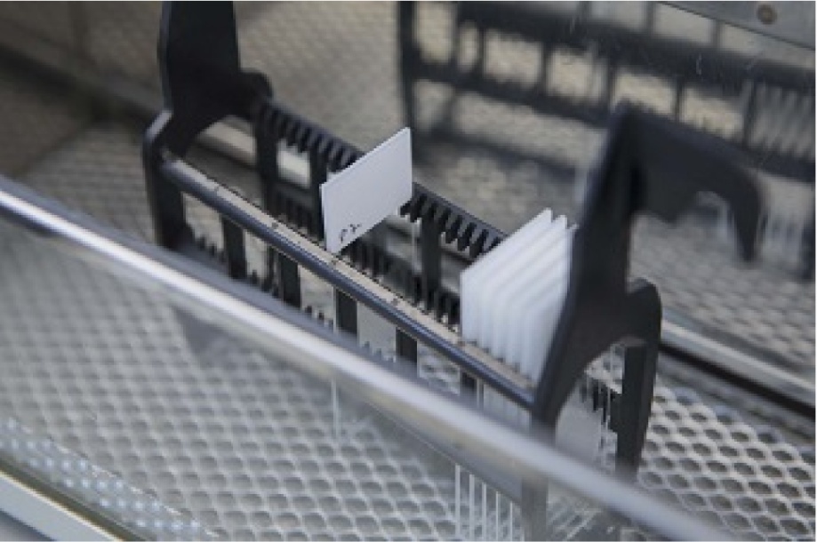
An overview of the different steps involved in PD-L1 IHC can be seen below. Briefly, samples are initially processed and mounted onto microscope slides. Sample sections are then stained for PD-L1 by using a commercial IHC assay with an automated staining platform. Stained samples are then quality control checked by a trained pathologist.

Steps performed during PD-L1 testing

1.
Sections cut
Step 1 in PD-L1 testing - Sections cut
2.
Sections mounted
Step 2 in PD-L1 testing - Sections mounted
3.
Slides dried
Step 3 in PD-L1 testing - Slides dried
4.
Slides labelled
Step 4 in PD-L1 testing - Slides labelled
5.
Automated IHC stainer
Step 5 in PD-L1 testing - Automated IHC stainer
6.
Staining quality checked
Step 6 in PD-L1 testing - Staining quality checked

ABBREVIATION:
IHC, immunohistochemistry

Before PD-L1 expression can be assessed, samples must be appropriately processed in line with the recommended PD-L1 IHC assay recommendations.

Standard tissue sample preparation and processing procedure for PD-L1 testing*28,57–59

Formalin fixation

  • 10% (v/v) neutral-buffered formalin
  • Optimal fixation times vary between assays

Paraffin embedding

  • Embed tumour sample in a paraffin block

Sectioning

  • Cut 4–5µm sections onto microscope slides
  • Dry slides at 58 ± 2ºC for 1 hour
  • Store at 2–8ºC or room temperature, to protect from light

*The preparation instructions differ for each PD-L1 assay. For further details, refer to specific package insert for individual assays.

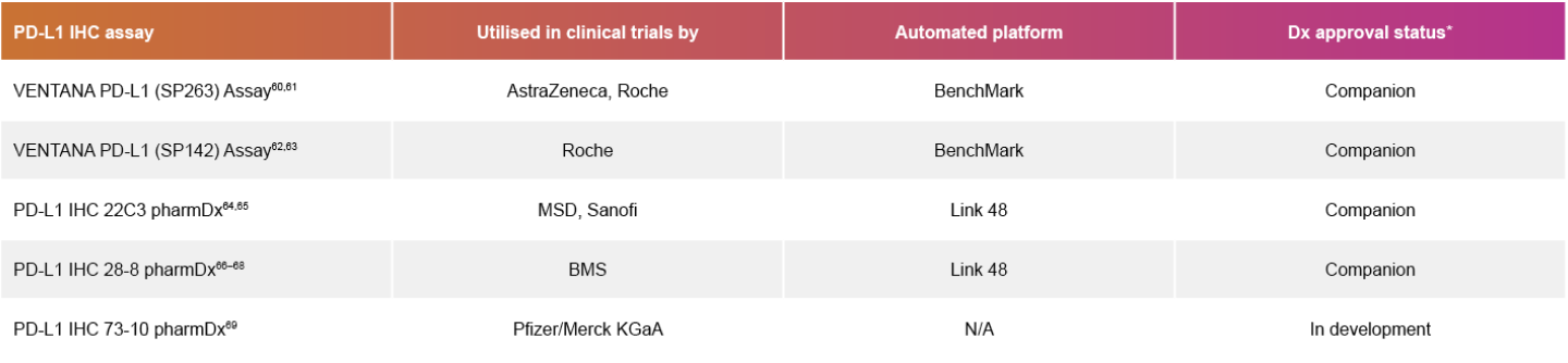
There are five IHC assays, either commercially available or in development, which can be used to determine PD-L1 expression. The choice of PD-L1 IHC assay and the staining procedure is used in line with specific immunotherapies according to their respective diagnostic labels.
Table of specific immunotherapies according to their respective diagnostic labelsTable of specific immunotherapies according to their respective diagnostic labels

*Based on the FDA and CE-IVD diagnostic regulatory approval51

ABBREVIATIONS:
BMS, Bristol Myers Squibb; CE-IVD, Conformité Européenne-in vitro diagnostic; Dx, diagnostic; FDA, Food and Drug Administration; IHC, immunohistochemistry; MSD, Merck Sharp & Dohme; N/A, not applicable; PD-L1, programmed cell death ligand-1

An LDT is an alternative approach for those with limited or no access to commercially developed PD-L1 assays. In this regard, LDTs are PD-L1 IHC assays that contain components from a variety of sources and apply custom procedures (de-novo LDT); or are based on the components from a regulatory-approved assay, but either apply a custom procedure to detect PD-L1 or for a different purpose (kit-derived LDT).70

The use of LDTs is associated with less reliable capture of PD-L1 positivity in NSCLC samples, when compared with approved PD-L1 assays.71,72

It is important that LDTs are adequately validated before their use on patient samples.70

ABBREVIATIONS:
ALK, anaplastic lymphoma kinase; BRAF, B-Raf proto-oncogene; CDx, companion diagnostic; CE-IVD, Conformité Européenne-in vitro diagnostic; ctDNA, circulating tumour DNA; Dx, diagnostic; (EBUS)-TBNA, (endobronchial ultrasound-)guided transbronchial needle aspiration; EGFR, epidermal growth factor receptor; EUS-FNA, endoscopic ultrasound-guided fine needle aspiration; FDA, US Food and Drug Administration; FFPE, formalin-fixed, paraffin-embedded; FISH, fluorescent in situ hybridisation; HER2, human epidermal growth factor receptor 2; IHC, immunohistochemistry; KRAS, Kirsten rat sarcoma virus; LDT, laboratory-developed test; MET, MET proto-oncogene, receptor tyrosine kinase; NGS, next-generation sequencing; NSCLC, non-small cell lung cancer; NTRK1/2/3, neurotrophic receptor tyrosine kinase 1/2/3; PCR, polymerase chain reaction; PD-L1, programmed cell death ligand-1; RET, rearranged during transfection; RGQ, Rotor-Gene Q; ROS1, ROS proto-oncogene 1; RT-PCR, real-time polymerase chain reaction; VATS, video-assisted thoracoscopic surgery

REFERENCES:

  1. Hendriks LE, et al. Non-oncogene-addicted metastatic non-small-cell lung cancer: ESMO Clinical Practice Guideline for diagnosis, treatment and follow-up. Ann Oncol 2023;34:358–76.
  2. Hendriks LE, et al. Oncogene-addicted metastatic non-small-cell lung cancer: ESMO Clinical Practice Guideline for diagnosis, treatment and follow-up. Ann Oncol 2023;34:339–57.
  3. Foundation Medicine. FoundationOne®CDx Technical Information. Available at: https://www.accessdata.fda.gov/cdrh_docs/pdf17/P170019S011C.pdf (accessed January 2024).
  4. Medicine. FoundationOne® Liquid CDx Technical Information. Available at: https://www.foundationmedicine.com/sites/default/files/media/documents/2023-11/P190032_S011_F1LCDx%20Technical%20Label%5B76%5D.pdf (accessed January 2024).
  5. Jenkins S, et al. Plasma ctDNA analysis for detection of the EGFR T790M mutation in patients with advanced non-small cell lung cancer. J Thorac Oncol 2017;12:1061–70.
  6. Ellison G, et al. EGFR mutation testing in lung cancer: a review of available methods and their use for analysis of tumour tissue and cytology samples. J Clin Pathol 2013;66:79–89.
  7. Wang, S, et al. The suitability of small biopsy and cytology specimens for EGFR and other mutation testing in non-small cell lung cancer. Transl Lung Cancer Res 2015;4:119–25.
  8. Oxnard GR, et al. Association between plasma genotyping and outcomes of treatment with osimertinib (AZD9291) in advanced non-small-cell lung cancer. J Clin Oncol 2016;34:3375–82.
  9. Du X, et al. ALK-rearrangement in non-small-cell lung cancer (NSCLC). Thorac Cancer 2018;9:423–30.
  10. Minca EC, et al. ALK status testing in non-small-cell lung carcinoma by FISH on ThinPrep slides with cytology material. J Thorac Oncol 2014;9:464–8.
  11. Lindeman NI, et al. Updated molecular testing guideline for the selection of lung cancer patients for treatment with targeted tyrosine kinase inhibitors: guideline from the College of American Pathologists, the International Association for the Study of Lung Cancer, and the Association for Molecular Pathology. Arch Pathol Lab Med 2018;142:321–46.
  12. Mezquita L, et al. An amplicon-based liquid biopsy for detecting ALK and ROS1 fusions and resistance mutations in advanced non-small cell lung cancer (NSCLC) patients. J Clin Oncol 2018;36(15_Suppl.):9095.
  13. NeoGenomics Laboratories. InVision First Lung. Available at: https://neogenomics.com/invisionfirstr-lung (accessed January 2024).
  14. McCoach CE, et al. Clinical utility of cell-free DNA for the detection of ALK fusions and genomic mechanisms of ALK inhibitor resistance in non-small cell lung cancer. Clin Cancer Res 2018;24:2758–70.
  15. Zhang L, et al. High feasibility of cytological specimens for detection of ROS1 fusion by reverse transcriptase PCR in Chinese patients with advanced non-small-cell lung cancer. OncoTargets and Ther 2019;12:3305–11.
  16. Viola P, et al. A validation study for the use of ROS1 immunohistochemical staining in screening for ROS1 translocations in lung cancer. J Thorac Oncol 2016;11:1029–39.
  17. Lin Q, et al. The association between BRAF mutation class and clinical features in BRAF-mutant Chinese non-small cell lung cancer patients. J Transl Med 2019;17:298.
  18. Brustugun OT, et al. BRAF-mutations in non-small cell lung cancer. Lung Cancer 2014;84(1):36–8.
  19. Qiagen. therascreen® KRAS RGQ PCR Kit Instructions for Use (Handbook). Available at: https://www.accessdata.fda.gov/cdrh_docs/pdf11/P110027S012C.pdf (accessed January 2024).
  20. Wu HH, et al. Utilization of cell-transferred cytologic smears in detection of EGFR and KRAS mutation on adenocarcinoma of lung. Mod Pathol 2014;27:930–5.
  21. Pruis MA, et al. Highly accurate DNA-based detection and treatment results of MET exon 14 skipping mutations in lung cancer. Lung Cancer 2020;140:46–54.
  22. Reale ML, et al. Molecular Testing and Treatment Strategies in RET-Rearranged NSCLC Patients: Stay on Target to Look Forward. J Mol Pathol 2022;3:24–37.
  23. Li T, et al. Advances in the Diagnosis and Treatment of a Driving Target: RET Rearrangements in non-Small-Cell Lung Cancer (NSCLC) Especially in China. Tech Cancer Res Treat 2023;22:153303382211488.
  24. Ren S, et al. Consensus for HER2 alterations testing in non-small-cell lung cancer. ESMO Open 2022;7:100395.
  25. Hofman P. What Is New in Biomarker Testing at Diagnosis of Advanced Non-Squamous Non-Small Cell Lung Carcinoma? Implications for Cytology and Liquid Biopsy. J Mol Pathol 2021;2:147–72.
  26. Reck M, et al. Pembrolizumab versus chemotherapy for PD-L1-positive non-small-cell lung cancer. N Engl J Med 2016;375:1823–33.
  27. Heymann JJ, et al. PD-L1 expression in non-small cell lung carcinoma: Comparison among cytology, small biopsy, and surgical resection specimens. Cancer Cytopathol 2017;125:896–907.
  28. Roche. VENTANA PD-L1 (SP263) Assay package insert. Available from: https://elabdoc-prod.roche.com/eLD/api/downloads/4b0ce998-2fb6-ea11-fc90-005056a71a5d?countryIsoCode=gb (accessed January 2024).
  29. Referenced with permission from the NCCN Clinical Practice Guidelines in Oncology (NCCN Guidelines®) for Non-Small Cell Lung Cancer V.11.2024. © National Comprehensive Cancer Network, Inc. 2024. All rights reserved. Accessed October 28, 2024. To view the most recent and complete version of the guideline, go online to NCCN.org. NCCN makes no warranties of any kind whatsoever regarding their content, use or application and disclaims any responsibility for their application or use in any way.
  30. Kim H, Chung JH. PD-L1 testing in non-small cell lung cancer: past, present, and future. J Pathol Transl Med 2019;53:199–206.
  31. Midha A, et al. PD-L1 expression in advanced NSCLC: primary lesions versus metastatic sites and impact of sample age. J Clin Oncol 2016;34(15_Suppl.):3025.
  32. Gagné A, et al. Impact of specimen characteristics on PD-L1 testing in non-small cell lung cancer: validation of the IASLC PD-L1 testing recommendations. J Thorac Oncol 2019;14:2062–70.
  33. Munari E, et al. PD-L1 expression comparison between primary and relapsed non-small cell lung carcinoma using whole sections and clone SP263. Oncotarget 2018;9:30465–71.
  34. Ofiara LM, et al. Optimizing Tissue Sampling for the Diagnosis, Subtyping, and Molecular Analysis of Lung Cancer. Front Oncol 2014;4:253.
  35. Lyng H, et al. GeneCount: genome-wide calculation of absolute tumor DNA copy numbers from array comparative genomic hybridization data. Genome Biol 2008;9:R86.
  36. Khalifa E, et al. Idylla EGFR assay on extracted DNA: advantages, limits and place in molecular screening according to the latest guidelines for non-small-cell lung cancer (NSCLC) patients. J Clin Pathol 2023;76:698–704.
  1. Cho M, et al. Tissue recommendations for precision cancer therapy using next generation sequencing: a comprehensive single cancer center’s experiences. Oncotarget 2017;8:42478–86.
  2. Lozano MD, et al., Cytology smears in the era of molecular biomarkers in non-small cell lung cancer: doing more with less. Arch Pathol Lab Med 2018;142:291–8.
  3. Schwaederle M, et al. Detection rate of actionable mutations in diverse cancers using a biopsy-free (blood) circulating tumor cell DNA assay. Oncotarget 2016;7:9707–17.
  4. Santarpia M, et al. Liquid biopsies to optimize therapeutic efficacy in unresponsive lung cancer patients. Expert Opin Drug Metab Toxicol 2018;14:761–3.
  5. Rolfo C, et al. Liquid Biopsy for Advanced Non-Small Cell Lung Cancer (NSCLC): A Statement Paper from the IASLC. J Thorac Oncol 2018;13:1248–68.
  6. Zito Marino F, et al. Molecular heterogeneity in lung cancer: from mechanisms of origin to clinical implications. Int J Med Sci 2019;16:981–89.
  7. Elazezy M, Joosse SA. Techniques of using circulating tumor DNA as a liquid biopsy component in cancer management. Comput Strut Biotechnol J 2018;16:370–8.
  8. MayoClinic. Video-assisted thoracoscopic surgery (VATS). Available at: https://www.mayoclinic.org/tests-procedures/video-assisted-thoracic-surgery/about/pac-20384922 (accessed January 2024).
  9. Patient.Info. Mediastinoscopy. Available at: https://patient.info/treatment-medication/mediastinoscopy (accessed January 2024).
  10. Cancer.gov. Bronchial brushing. Available at: https://www.cancer.gov/publications/dictionaries/cancer-terms/def/bronchial-brushing (accessed January 2024).
  11. Cancer.gov. EUS-FNA. 2024 Available at: https://www.cancer.gov/publications/dictionaries/cancer-terms/def/eus-fna (accessed January 2024).
  12. Patient Info. Endobronchial ultrasound-guided transbronchial needle aspiration. Available from:https://patient.info/treatment-medication/endobronchial-ultrasound-guided-transbronchial-needle-aspiration (accessed January 2024).
  13. Goto T, et al. Distribution of circulating tumor DNA in lung cancer: analysis of the primary lung and bone marrow along with the pulmonary venous and peripheral blood. Oncotarget 2017;8:59268–81.
  14. Pennell NA, et al. Biomarker testing for patients with advanced non-small cell lung cancer: real-world issues and tough choices. Am Soc Clin Oncol Educ Book 2019;39:531–42.
  15. FDA. List of Cleared or Approved Companion Diagnostic Devices (In Vitro and Imaging Tools). Available from:https://www.fda.gov/medical-devices/vitro-diagnostics/list-cleared-or-approved-companion-diagnostic-devices-vitro-and-imaging-tools (accessed January 2024).
  16. Qiagen. therascreen® EGFR RGQ PCR Kit Handbook. Available from: https://www.qiagen.com/au/resources/resourcedetail?id=394a7dca-988a-4453-a399-99f55d67c2f5&lang=en (accessed January 2024).
  17. Roche. cobas® EGFR Mutation Test v2. Available from: https://elabdoc-prod.roche.com/eLD/api/downloads/71bf89fb-e86d-ee11-2091-005056a71a5d?countryIsoCode=gb (accessed January 2024).
  18. Huber D, et al. Fluorescence in situ hybridization (FISH): history, limitations and what to expect from micro-scale FISH? Micro Nano Eng 2018;1:15–24.
  19. Kim SW, et al. Immunohistochemistry for pathologists: protocols, pitfalls, and tips. J Pathol Transl Med 2016;50:411–18.
  20. Isla D, et al. New update to the guidelines on testing predictive biomarkers in non-small-cell lung cancer: a National Consensus of the Spanish Society of Pathology and the Spanish Society of Medical Oncology. Clin Transl Oncol 2023;25:1252–67.
  21. Roche. VENTANA PD-L1 (SP142) Assay package insert (FDA). Available at: https://www.accessdata.fda.gov/cdrh_docs/pdf16/p160002c.pdf (accessed January 2024).
  22. Dako. PD-L1 IHC 28-8 pharmDx package insert (FDA). Available at: https://www.accessdata.fda.gov/cdrh_docs/pdf15/P150025S003C.pdf (accessed January 2024).
  23. Dako. PD-L1 IHC 22C3 pharmDx package insert (FDA). Available at: https://www.agilent.com/cs/library/packageinsert/public/P03951E_20.pdf (accessed January 2024).
  24. Roche. VENTANA PD-L1 (SP263) Assay (CE IVD). Available at: https://diagnostics.roche.com/gb/en/products/tests/ventana-pd-l1-_sp263-assay2.html#productSpecs (accessed January 2024).
  25. Roche. VENTANA PD-L1 (SP263) Assay (US-FDA). Available at: https://diagnostics.roche.com/global/en/products/lab/pd-l1-sp263-assay-ventana-rtd001235.html (accessed January 2024).
  26. Roche. VENTANA PD-L1 (SP142) Assay (CE IVD). Available at: https://diagnostics.roche.com/global/en/products/lab/pd-l1-sp142-assay-us-export-ventana-rtd001232.html (accessed January 2024).
  27. Roche. VENTANA PD-L1 (SP142) Assay (US-FDA). Available at: https://diagnostics.roche.com/global/en/products/lab/pd-l1-sp142-assay-ventana-rtd001231.html (accessed January 2024).
  28. Agilent. PD-L1 IHC 22C3 pharmDx Overview (EU). Available at: https://www.agilent.com/en/product/pharmdx/pd-l1-ihc-22c3-pharmdx-overview (accessed January 2024).
  29. Agilent. PD-L1 IHC 22C3 pharmDx Overview (US). Available at: https://www.agilent.com/en-us/product/pharmdx/pd-l1-ihc-22c3-pharmdx-overview (accessed January 2024).
  30. Agilent. PD-L1 IHC 28-8 pharmDx for non-squamous NSCLC (US). Available at: https://www.agilent.com/en/product/pharmdx/pd-l1-ihc-28-8-pharmdx-for-non-squamous-nsclc (accessed January 2024).
  31. Agilent. PD-L1 IHC 28-8 pharmDx for non-squamous NSCLC (EU). Available from: https://www.agilent.com/en/product/pharmdx/pd-l1-ihc-28-8-pharmdx-for-non-squamous-nsclc (accessed January 2024).
  32. OncLive. FDA approves PD-L1 IHC 28-8 pharmDx as companion diagnostic for nivolumab plus ipilimumab in NSCLC. 2020 Available from: https://www.onclive.com/view/fda-approves-pdl1-ihc-288-pharmdx-as-companion-diagnostic-for-nivolumabipilimumab-in-nsclc (accessed January 2024).
  33. Patel MR, et al. Avelumab in metastatic urothelial carcinoma after platinum failure (JAVELIN Solid Tumor): pooled results from two expansion cohorts of an open-label, phase 1 trial. Lancet Oncol 2018;19:51–64.
  34. Cheung CC, et al. Fit-for-purpose PD-L1 biomarker testing for patient selection in immuno-oncology: guidelines for clinical laboratories from the Canadian Association of Pathologists-Association Canadienne Des Pathologistes (CAP-ACP). Appl Immunohistochem Mol Morphol 2019;27:699–714.
  35. Velcheti V, et al. Real-world PD-L1 testing and distribution of PD-L1 tumor expression by immunohistochemistry assay type among patients with metastatic non-small cell lung cancer in the United States. PloS ONE 2018;13:e0206370.
  36. Adam J, et al. Multicenter harmonization study for PD-L1 IHC testing in non-small-cell lung cancer. Ann Oncol 2018;29:953–58.

Welcome to the iD LUNG website

Access to the iD Lung website is for non-US healthcare professionals, and is of particular relevance for medical oncologists, pathologists and the surgical team.

By clicking on the link below, you declare and confirm that you are a non-US healthcare professional.

Access the site

Patients or members of the public

This website is intended for healthcare professionals only. Please speak to your healthcare provider if you require further information on lung cancer.

Media

If you are a member of the press, please contact the AstraZeneca Press Office.

Content

The content of this website is not exhaustive and should not be used as a substitute for the information provided by the manufacturers of the diagnostic assays. For specific guidance on testing protocols, please consult the package insert provided with each diagnostic test.

iD.Lung.com

  • Introduction
  • Biomarker Testing in Lung Cancer
  • Technical Considerations
  • iD Podcasts
  • Resources & Links
  • FAQs
  • Contact Us
  • Legal Notice
  • Privacy Policy
  • Cookies
AstraZeneca Logo

©AstraZeneca 2024
FOR HEALTHCARE PROFESSIONAL USE ONLY
Veeva Document ID: Z4-60052 | Date of preparation: September 2024